Eye Examination
What to Expect at an Eye Examination
The doctor will ask you a number of questions, regarding the purpose of your visit. Please bring a list of your medications and a detailed health history. Generally, most patients require about an hour for an examination. However, depending upon how long it takes for the eye drops to take effect and how many additional tests you require, sometimes you can be at our office as long as 2 hours. After the examination, your vision may be blurry and light sensitive for several hours. Please bring sunglasses. We can also provide you with disposable sunglasses. You may not feel comfortable to drive until the eye drops wear off.
During a comprehensive eye examination at our clinic, the following will be evaluated: Medical and Ocular case history, high-tech pretesting, visual acuity, color vision testing, refraction, intraocular pressure, ocular health & retinal assessment. Additional tests may be required depending upon risk factors and findings. Many eye diseases such a glaucoma, have no symptoms until irreversible damage has occurred. With routine examinations and the advent of improved ocular imaging, many diseases can be detected in the early stages and vision can be preserved with early intervention and treatment.
Hi-Tech Pre-testing
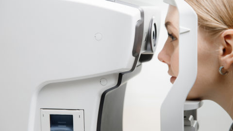
- Auto refraction (an estimate of refractive error)
- Auto keratometry (corneal curvature measurement)
- Intraocular eye pressure for glaucoma
- Pachymetry corneal thickness (non contact) for glaucoma and corneal assessment
Medical and Ocular Case History
- Detailed personal and family history of eye disease and medical conditions
- Current concerns and symptoms
Visual Acuity
- Ability to see with and without glasses at distance and near
Colour Vision Testing
- For children and as required in adults
Subjective Refraction
- Determine the prescriptions for eye glasses
Binocular Vision and Eye Muscles
- Eye muscle evaluation to determine if there is a misalignment of the eyes and how well the two eyes work together
- Stereo acuity (depth perception) for children and as required in adults
- Determine if prism is required
Pupillary Reflexes
- Can be abnormal if there is optic nerve or ocular motor damage or any abnormality along the visual pathway in the brain
Slit Lamp Bio Microscope
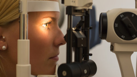
- Evaluation of the Cornea, Crystalline lens, Eye lids and Conjunctiva to determine the presence of cataracts, diseases of the eye lids, cornea and conjunctiva
- Determine the presence of inflammation in the front chamber of the eye
- Tear film evaluation and dry eye assessment
Retinal Examination
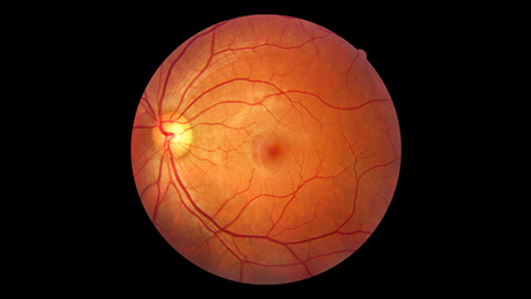
- In most cases, drops are put in the eyes to dilate the pupils and allow the doctor to examine the retina (the back of the eye). A large portion of the retina is not visible to the doctor, unless the pupils are dilated. A dilated examination of the retina is essential in order to find retinal tears, holes, detachments and other retinal diseases.
- The optic nerve and macula (central retina) are examined once the drops take effect.
- For an additional fee, retinal and optic nerve imaging will be performed and is strongly recommended
Additional Tests
Hi-Tech Imaging (Additional Fee)
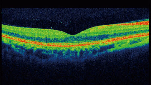
- Retinal imaging (OCT – Ocular Coherence Tomography) is strongly recommended to microscopically examine and measure the thickness of the layers of the macula (central retina), in order to detect early signs of macular degenerations, diabetic eye disease and many retinal disorders
- Optic nerve imaging (OCT – Ocular Coherence Tomography) is strongly recommend to determine early signs of glaucoma and other diseases of the optic nerve and some other neurological conditions.
- The thickness of the nerve fibre layer is measured and analyzed and glaucoma progression or stability can be determined.
Automated Visual Fields (Additional Fee)
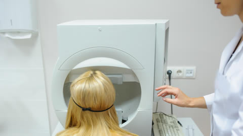
- Required if there are findings of risk factors for glaucoma during the the eye examinations or a family history of glaucoma
- Required if there is a history of headaches increasing in frequency or duration
- Required if there are any suspicious neurological findings or symptoms
- Required if there are certain retinal findings
- Required if there are stroke symptoms or a history of stroke
Gonioscopy
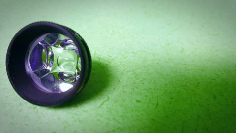
- Evaluation of the anterior chamber of the eye to determine if preventive laser treatment (Iridotomy) is require to prevent a narrow angle glaucoma attack
Summary of Findings and Treatment Plan
- Your eye doctor will summarize the findings of the examination and provide a treatment plan
- A referral to the appropriate healthcare practitioner will be arranged, if required
- A prescription for eye glasses will be provided
Contact Lens Evaluation
- Evaluate the current contact lenses fit and prescription
- Determine if a better lens option is available
- Determine if an adjustment in the Contact Lens parameters are required
- Determine the health of the ocular structures as relate to contact lens wear
Contact Lens Fitting
- Your eye doctor will determine the best fit and prescription to provide the best vision
- The technician will patiently teach you how to insert and remove your contacts
- A separate appointment is sometimes required for instruction on insertion and removal of contact lenses
Laser Surgery Co-Management
- Laser surgery pre-operative consultation and post operative care
- Extensive referral network of exceptional laser surgeons
Call 416-968-2300 to book an eye exam or visit our clinic

